Medicina Laboratorial e Patologia Clínica
Cowell and Tyler's Diagnostic Cytology and Hematology of the Dog and Cat, 6th Edition
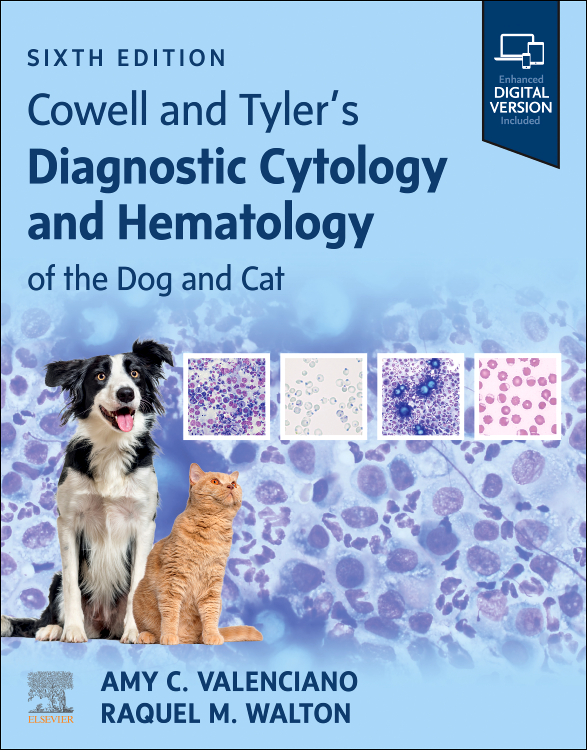
De: Amy C. Valenciano, Raquel Walton
ISBN: 9780443279515
2027, Elsevier
Capa dura
Páginas: 576
Medicina Laboratorial e Patologia Clínica
De: Amy C. Valenciano, Raquel Walton
ISBN: 9780443279515
2027, Elsevier
Capa dura
Páginas: 576
Gain important skills for accurate microscopic evaluation of blood, cytology specimens, and body fluids! Comprehensive yet practical, Cowell and Tyler's Diagnostic Cytology and Hematology of the Dog and Cat, 6th Edition, guides you through the clinical laboratory techniques required to diagnose a wide variety of pathologic conditions seen in everyday practice. Full-color photomicrographs demonstrate pertinent hematologic and cytologic features, help you identify many bacterial, fungal, and protozoal organisms, and differentiate normal cells from abnormal cells. Written by a team of experts, led by veterinary clinical pathologists Amy Valenciano and Raquel Walton, this trusted, widely used text is an ideal reference and guide to keep alongside the microscope.
Key Features
1. Sample Collection and Preparation
2. Cell Types and Criteria of Malignancy
3. Selected Infectious Agents
4. Round Cells
5. Cutaneous and Subcutaneous Lesions
6. Subcutaneous Glandular Tissue: Mammary, Salivary, Thyroid, and Parathyroid
7. Nasal Exudates and Masses
8. Oropharynx and Tonsils
9. Eyes and Associated Structures
10. External Ear Canal
11. The Lymph Nodes
12. Synovial Fluid Analysis
13. The Musculoskeletal System
14. Cerebrospinal Fluid and Central Nervous System Cytology
15. Effusions: Abdominal, Thoracic, and Pericardial
16. Transtracheal and Bronchoalveolar Washes
17. The Lung and Intrathoracic Structures
18. The Gastrointestinal Tract
19. The Pancreas
20. The Liver
21. The Spleen
22. The Kidneys
23. Examination of Urinary Sediment
24. The Male Reproductive Tract: Prostate, Testes, Penis, and Semen
25. The Female Reproductive Tract
26. Peripheral Blood Smears
27. The Bone Marrow
28. The Adrenal Gland
29. Immunocytochemistry
30. Flow Cytometry
31. Molecular Methods in Lymphoid Malignancies
Amy C. Valenciano, Veterinary Clinical Pathologist, IDEXX Cellular Diagnostics Research & Development, Dallas, Texas, USA
Raquel Walton, Veterinary Clinical Pathologist, IDEXX Laboratories, Inc., USA